
PRP in Dogs: Biological Principles, Clinical Application and Evidence
In brief
PRP derived from the patient's own blood has proven efficacy in joint osteoarthritis and tendon conditions – no allergy risk, combinable with hyaluronic acid, and adjuvant after joint surgery in dogs.
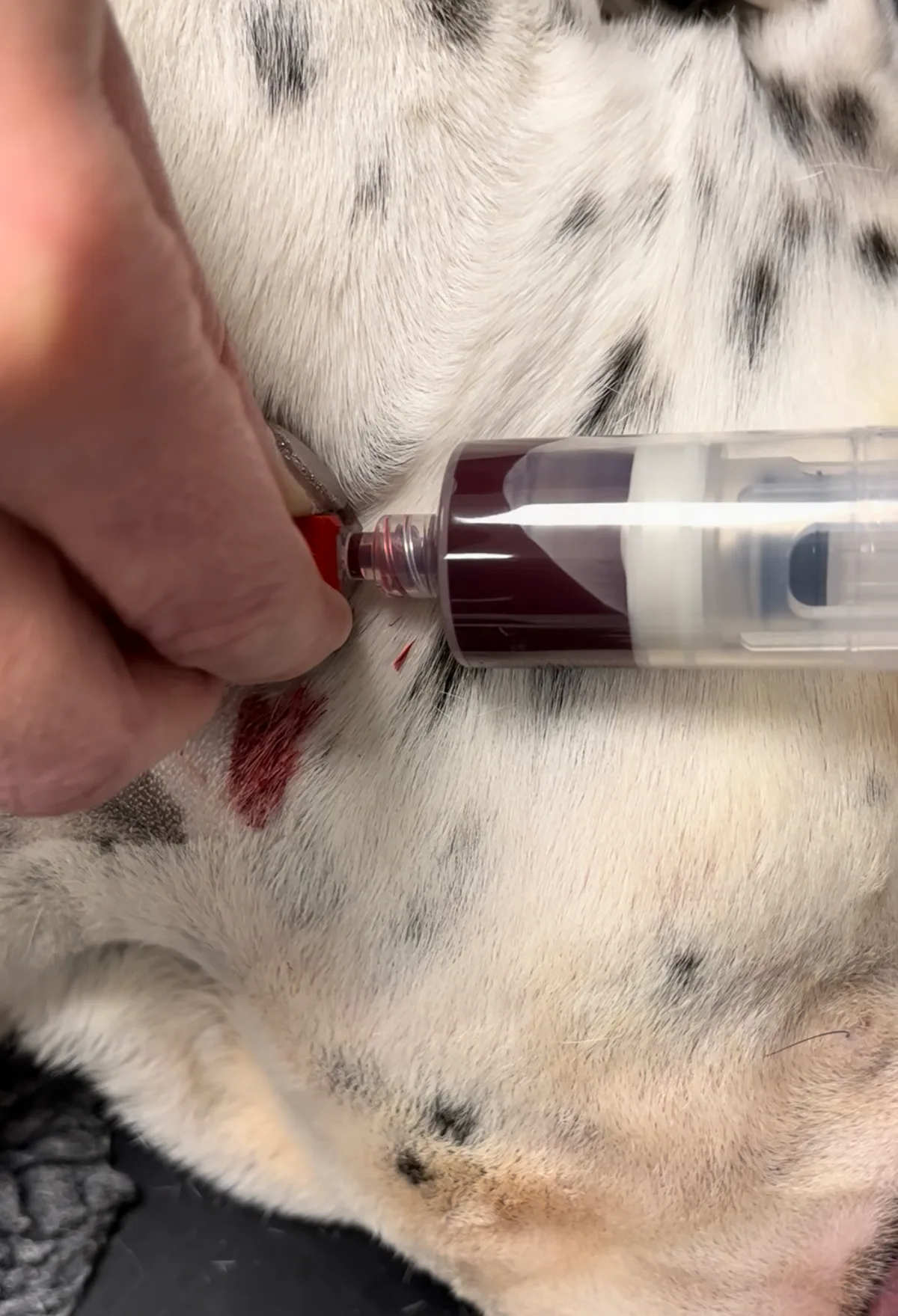
Platelet-rich plasma (PRP) has established itself in veterinary orthopaedics and surgery as a biological therapeutic with proven clinical benefit. Unlike conventional analgesics, PRP is not a drug in the pharmacological sense but an autologous blood product that contains concentrated growth factors and actively intervenes in the pathophysiology of joint degeneration, tendon healing and tissue repair.
Its use is multimodal: as a primary biological therapy in osteoarthritis and tendinopathy, and as a postoperative adjuvant to optimise tissue healing after joint surgery. This article explains the scientific principles, clinical indications and the critical quality requirements for PRP preparation.
What Is PRP – and Where Does It Come From?
PRP is a concentrated blood product prepared exclusively from the patient’s own blood – it is therefore completely autologous. From a blood sample, targeted centrifugation enriches the platelet fraction to a multiple of baseline values. These platelets are rich in growth factors that play a central role in tissue regeneration and inflammation regulation.
The key growth factors in PRP include:
- PDGF (Platelet-Derived Growth Factor) – promotes cell migration and proliferation
- TGF-β1/β2 (Transforming Growth Factor) – anti-inflammatory, promotes collagen synthesis
- VEGF (Vascular Endothelial Growth Factor) – stimulates new vessel formation
- bFGF (Basic Fibroblastic Growth Factor) – stimulates connective tissue healing
- EGF (Epidermal Growth Factor) – supports cell growth and wound healing
These factors act both individually and synergistically: they inhibit inflammatory cytokines, activate endogenous stem cells, promote chondrocytes and stimulate extracellular matrix synthesis. In short: PRP supports the body’s own healing processes at a biological level.
Indications in Dogs
Osteoarthritis
Osteoarthritis is the most common cause of chronic lameness in dogs and affects particularly older and large-breed animals. Several clinical studies confirm that intraarticular PRP injections in dogs can reduce lameness and objectively improve weight bearing on the affected limb.
A robust study by Fahie et al. (2013) showed that autologous platelet concentrate led to significant clinical improvement in dogs with osteoarthritis. Vilar et al. (2018) documented with gait analysis a measurable increase in loading force after four injections in dogs with stifle osteoarthritis following cruciate rupture – the effect was already demonstrable at 30 days. A duration of effect of 3–6 months is consistent with the current clinical and scientific evidence.
Cruciate Ligament Pathology (Partial Rupture / Instability)
Cook et al. (2016) showed in dogs with partial cruciate rupture and meniscal lesion that multiple injections of leucoreduced PRP improved range of motion, reduced pain and improved function for up to six months – with a measurable effect already one week after the first injection.
Tendon Conditions: Supraspinatus, Biceps and Others
Tendon conditions are common in sporting, agility and working dogs. The supraspinatus tendon and biceps tendon are particularly affected by degenerative changes (tendinopathy). PRP is administered here under ultrasound guidance intraparatendinously.
Canapp et al. (2016) published a study of 55 dogs treated for supraspinatus tendinopathy with a combination of adipose-derived progenitor cells (ADPC) and PRP. In 82% of cases, the cross-sectional area of the affected tendon reduced to the size of the healthy tendon within 90 days – with a significantly improved fibre pattern on ultrasound. PRP offers a meaningful advantage over conservative therapy here: it does not merely target symptom control but actively promotes structural tissue healing.
PRP as a Surgical Adjuvant
PRP combines excellently with surgical procedures. Silva et al. (2013) injected autologous platelet concentrate postoperatively after cruciate surgery and demonstrated significantly better gait outcomes in the PRP group at 90 days postoperatively. PRP appears to accelerate tissue healing after surgery and slow arthritis development – both clinically relevant benefits after TPLO, for example.
PRP + Hyaluronic Acid: More Than the Sum of Its Parts
A particularly promising strategy is the combination of PRP with hyaluronic acid (HA). The meta-analysis by Aw et al. (2021, Journal of Experimental Orthopaedics) encompassed 10 studies with 983 patients and showed that the PRP + HA combination consistently delivers better outcomes than PRP alone.
The synergy can be explained biologically: HA improves the joint environment through lubrication and matrix protection, dampens the transient inflammatory reaction (“post-injection flare”) that can follow PRP injection, and protects chondrocytes from oxidative stress and proteolytic enzymes. We therefore routinely offer the PRP-HA combination.
Not All PRP Is Equal – and This Makes a Critical Difference
PRP quality depends substantially on the preparation method. A central distinguishing characteristic is leucocyte content:
Leucocyte-poor PRP (LP-PRP) contains few neutrophils and red blood cells. It is considered the preferred product for intraarticular and intratendinous injections, because neutrophils release pro-inflammatory mediators (IL-1β, IL-6, TNF-α) and activate metalloproteinases (MMP-9) that degrade collagen and cartilage matrix.
Leucocyte-rich PRP (LR-PRP) contains substantially more neutrophils. In-vitro studies show that LR-PRP causes significantly more synoviocyte cell death than LP-PRP – an undesirable effect, particularly in an already damaged joint.
The method we use achieves a platelet enrichment of 500–550% relative to whole blood, with a simultaneous reduction in neutrophils of more than 85%. A 4–7-fold platelet enrichment corresponds to the target range established in human medicine.
PRP in Dogs vs. Librela – Safety Profile and Clinical Reality
Librela (bedinvetmab) is a monoclonal anti-NGF antibody that has been used as a monthly pain therapy for osteoarthritis since its approval. However, increasingly serious adverse events have been reported, including catastrophic damage to osteoarthritic joints. Furthermore, Librela is a purely analgesic therapy with no demonstrated disease-modifying or regenerative effect.
PRP, by contrast, is completely autologous: because it is derived from the patient’s own blood, immune-mediated reactions, foreign-protein-associated side effects and systemic toxicity are excluded by definition. Adverse effects are limited to a brief local inflammatory reaction (post-injection flare) within 24–72 hours, which is self-limiting. PRP additionally has the potential not only to relieve pain but to actively promote tissue repair processes.
On the basis of current safety data, we advise against the routine use of Librela as first-line therapy. In particular, the risk of rapidly progressive destructive joint changes (Rapidly Progressive Osteoarthritis, RPOA) – well documented in human medicine under anti-NGF therapies and increasingly discussed in the veterinary context – represents a substantial safety concern.
What Does Treatment Involve?
Treatment is performed under brief sedation or short general anaesthesia:
- Blood collection
- Two-step centrifugation for platelet enrichment (LP-PRP protocol)
- Quality control of platelet concentration (target: 4–7-fold enrichment)
- Injection into the joint (intraarticular) or tendon (intratendinous, with ultrasound guidance where indicated)
- Rest for 48 hours, then controlled gradual increase in activity
For osteoarthritis treatment we typically recommend a series of 2–3 injections at 4–6-week intervals, frequently combined with hyaluronic acid. The effect usually begins within 1–4 weeks and lasts 3–6 months. Treatment can be repeated as needed.
Preparation: Anti-inflammatory medications do not need to be stopped. Any corticosteroid therapy should be discontinued at least 1 week before treatment begins. Your dog must be fasted for the required sedation.
Frequently Asked Questions
Is PRP safe for dogs? Yes. Because PRP is derived from the patient’s own blood, there is no risk of allergic or rejection reactions. Side effects are limited in rare cases to a brief post-injection inflammatory reaction (injection flare) that resolves within 1–3 days.
Can PRP replace surgery? Not in all cases. For complete cruciate ligament rupture, surgery (e.g. TPLO) remains the treatment of choice. However, PRP can be used as an adjuvant to accelerate healing and slow the progression of osteoarthritis.
Conclusion
PRP is not a miracle therapy – but a well-supported, safe and versatile biological option that, when correctly prepared and appropriately applied, offers genuine clinical value. The quality of the product is decisive: not every system marketed as PRP actually delivers a platelet-enriched preparation. We use a validated leucocyte-poor PRP protocol in line with current scientific evidence.
If your dog suffers from osteoarthritis, tendon conditions or cruciate ligament pathology, please get in touch – we will advise you individually on whether PRP is a suitable option.