Skip to content
OsteoarthritisLibrelaSolensia

Librela – Wonderdrug or hidden risk?

In brief

Librela (bedinvetmab) is a monoclonal antibody against nerve growth factor licensed for canine osteoarthritis pain – and similarly Solensia in cats. This article reviews the growing pharmacovigilance evidence and positions Librela safely within the OA treatment cascade.

A note on Librela® (bedinvetmab) — Adverse effects & safe positioning in the osteoarthritis treatment cascade

Whilst the benefits of Librela in treating osteoarthritis-associated pain are undeniable, this article aims to raise awareness of its increasingly frequently reported musculoskeletal side-effects.

Bedinvetmab is a monoclonal antibody against nerve growth factor (NGF), licensed for the alleviation of pain associated with canine osteoarthritis (OA) — similarly to Solensia® in cats. Amongst other roles, NGF is involved in nociception and bone remodelling.

Human anti-NGF experience

Anti-NGF monoclonal antibodies (e.g. tanezumab, Pfizer) in humans were associated with increased rates of Rapidly Progressive Osteoarthritis (RPOA) and joint destruction. No anti-NGF therapy has been granted FDA (USA) or EMA (European) approval for osteoarthritis pain in humans, due to joint-safety concerns and an unfavourable risk–benefit balance. This represents a class safety issue, not an isolated molecule issue.

Rapidly Progressive Osteoarthritis (RPOA) is defined in human medicine as:

  • Accelerated joint destruction occurring over months, far exceeding the expected natural progression of OA
  • Characterised by rapid cartilage loss, subchondral bone collapse, fractures, luxation, and severe joint instability
  • These changes are irreversible once established

What was not adequately tested before Librela® rollout in dogs

(Farrell et al., Vet J 2024)

  • No large-scale radiographic screening for accelerated joint degeneration was performed in pivotal field trials
  • Only one small laboratory safety study assessed joint changes radiographically (limited numbers, short duration)
  • Fewer than 100 dogs received more than 3 monthly doses before market authorisation
  • Dogs were healthy, without naturally occurring OA (laboratory beagles); despite this, cartilage abnormalities were reported on post-mortem histology
  • Regulatory review acknowledged that a treatment-related contribution to bony/cartilage changes could not be excluded

Post-authorisation pharmacovigilance

Reported adverse events include: rapid joint destruction, pathological fractures, joint luxation, severe polyarthritis, neurological dysfunction, and euthanasia or death following musculoskeletal collapse.

Expert review of reported cases found multiple dogs with changes consistent with accelerated joint failure — sometimes months after treatment initiation — and judged the association as highly suspicious.

Of 4,746 reported musculoskeletal adverse events (MSAE) since Librela’s launch, 789 were filtered by independent investigators as pure MSAE (not confounded by neurological or systemic events). Compared to other medications with similar indications, Librela shows a 9-fold higher occurrence of MSAEs.
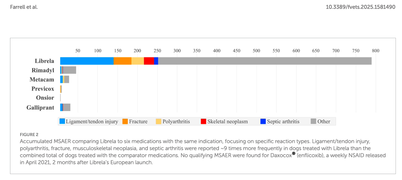
Bar chart: Accumulated musculoskeletal adverse events (MSAE) comparing Librela to six medications with the same indication. Librela shows a 9-fold higher MSAE rate than all comparator medications combined.
Figure 1. Accumulated musculoskeletal adverse events (MSAE) comparing Librela against six medications with the same indication (Rimadyl, Metacam, Previcox, Onsior, Galliprant), broken down by reaction type (ligament/tendon injury, fracture, polyarthritis, skeletal neoplasm, septic arthritis). Ligament/tendon injury, polyarthritis, fracture, musculoskeletal neoplasia, and septic arthritis were reported approximately 9 times more frequently in dogs treated with Librela than in the combined total of all comparator medications. No qualifying MSAE were found for Daxocox® (enflicoxib). Source: Farrell M et al. Frontiers in Veterinary Science 2025; doi: 10.3389/fvets.2025.1581490

Radiographic and CT findings

The following imaging documents cases of accelerated joint destruction under Librela therapy. The images speak for themselves.
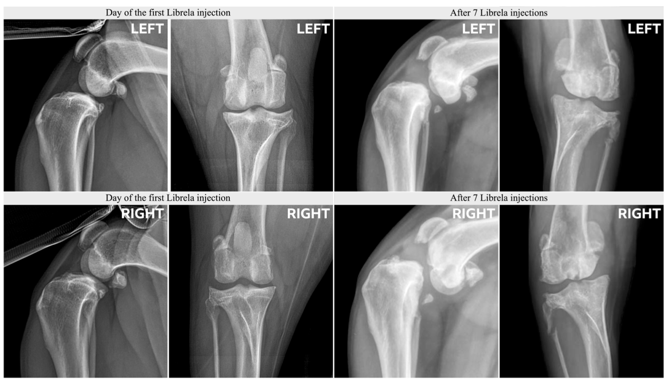
Radiographs of both stifle joints: on the day of the first Librela injection versus after 7 injections. Marked bilateral progression of joint destruction.
Figure 2. Mediolateral and caudocranial radiographs of both stifle joints. Left: Findings on the day of the first Librela injection — moderate degenerative joint changes. Right: Findings after 7 monthly Librela injections — marked progression of joint destruction with loss of normal joint architecture, periarticular new bone formation, and flattening of articular surfaces bilaterally. The rate of change far exceeds the expected natural progression of OA over this period. Source: Farrell M et al. Frontiers in Veterinary Science 2025; doi: 10.3389/fvets.2025.1581490
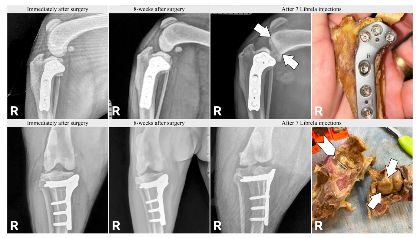
Radiographs and gross pathology of a dog after TPLO surgery: immediately post-operative, 8 weeks post-op, and after 7 Librela injections. Arrows indicate bone destruction. Gross pathology specimen shows the severity of joint destruction.
Figure 3. Radiographic progression and intraoperative gross pathology in a dog that had undergone TPLO (tibial plateau levelling osteotomy). Column 1: Immediately post-operatively — correct implant position, normal healing signs. Column 2: 8 weeks post-operatively — unremarkable healing progression. Column 3: After 7 monthly Librela injections — arrows indicate new subchondral bone destruction and periarticular lysis not present at 8 weeks. Column 4: Gross pathology specimen at revision surgery — extensive cartilage and bone destruction, fragmentation of the articular surface. Librela was commenced after TPLO; this case illustrates why Librela must not be used for post-operative pain management. Source: Farrell M et al. Frontiers in Veterinary Science 2025; doi: 10.3389/fvets.2025.1581490
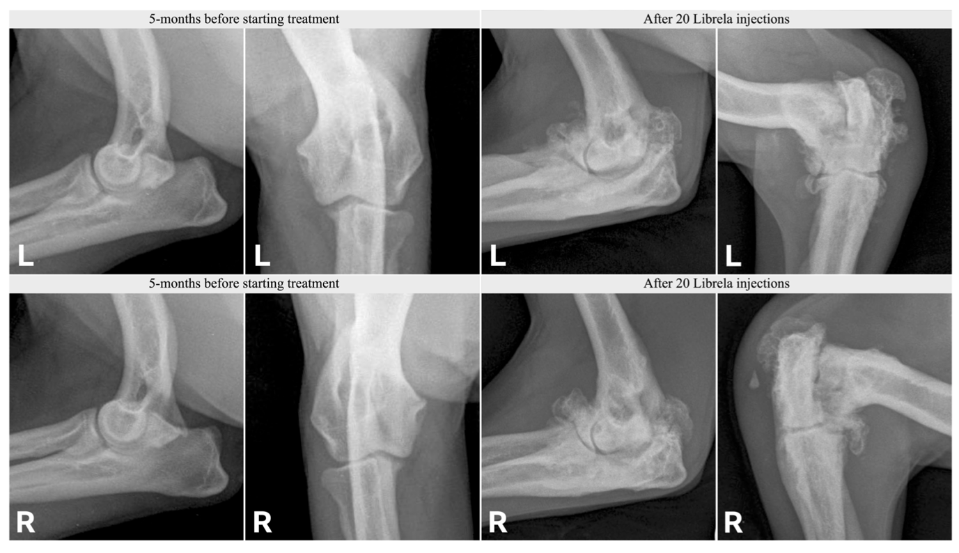
Radiographs of both elbow joints: 5 months before treatment versus after 20 Librela injections. Massive osteophyte formation and complete loss of normal joint architecture bilaterally.
Figure 4. Mediolateral and craniolateral radiographs of both elbow joints. Left (L and R): 5 months before starting Librela — moderate medial compartment disease as the baseline. Right (L and R): After 20 monthly Librela injections — massive periarticular new bone formation, complete loss of normal joint architecture, and signs of ankylosis with extensive osteophytosis bilaterally. The degree of progression over this period is inconsistent with the expected natural course of OA. Source: Farrell M et al. Frontiers in Veterinary Science 2025; doi: 10.3389/fvets.2025.1581490
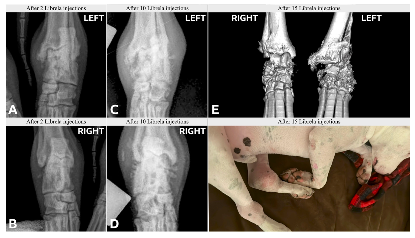
CT and radiographic images of the carpal joints: serial progression from 2 to 15 Librela injections. Clinical photograph shows massive bilateral soft-tissue swelling and deformity of the distal limbs.
Figure 5. Serial imaging of the carpal joints under Librela therapy. A/B (after 2 injections): Radiographs of left (A) and right (B) carpus — moderate periarticular changes. C/D (after 10 injections): Radiographs of left (C) and right (D) carpus — marked increase in destruction with loss of carpal bone definition. E (after 15 injections): Bilateral 3D CT reconstruction — complete collapse of the carpal bones, extensive periarticular mineralisation, and gross deformity of the distal limbs. Clinical photograph: massive bilateral soft-tissue swelling and deformity with apparent pain-free weight-bearing, illustrating how analgesia can mask the clinical signs of progressive structural destruction. Source: Farrell M et al. Frontiers in Veterinary Science 2025; doi: 10.3389/fvets.2025.1581490
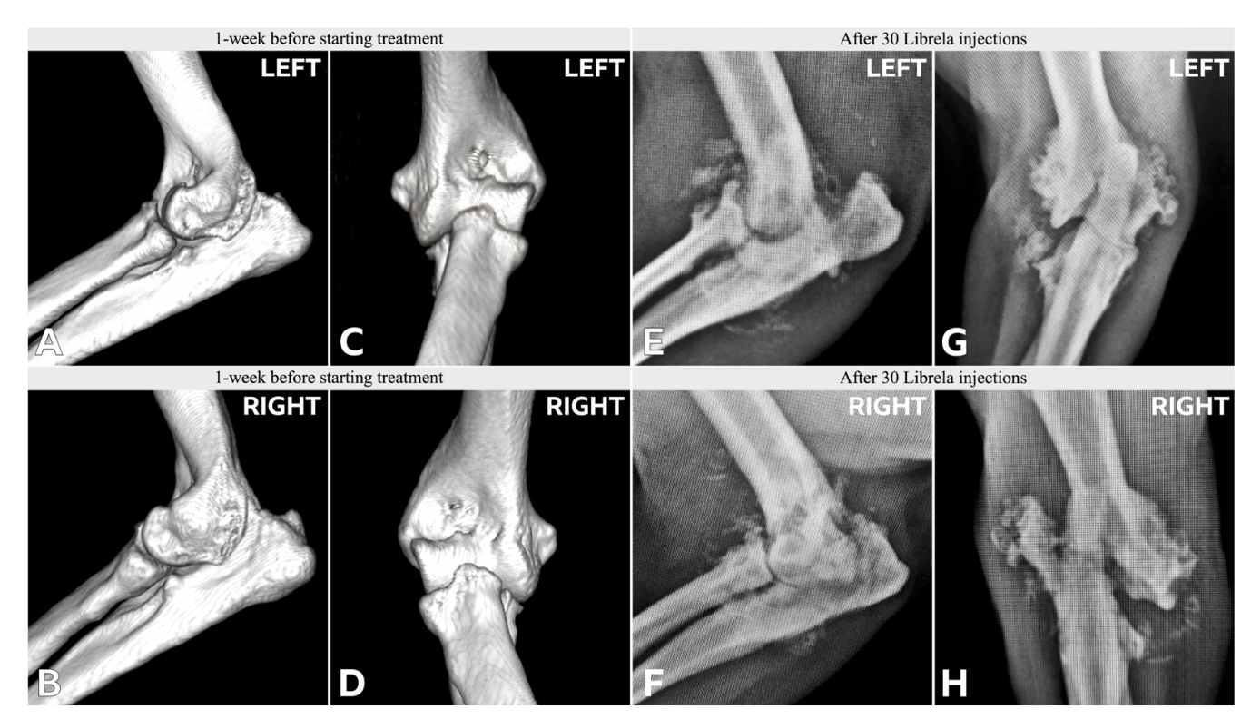
CT and radiographic images of the tarsal joints: 1 week before treatment versus after 30 Librela injections. Complete bilateral destruction of articular surfaces with extensive periarticular new bone.
Figure 6. Imaging of the tarsal (hock) joints before and after Librela therapy. A–D (1 week before starting treatment): 3D CT reconstructions (A/B) and radiographs (C/D) of both tarsal joints — moderate to advanced degenerative changes as baseline. E–H (after 30 monthly Librela injections): Radiographs (E/F) and CT (G/H) — complete destruction of articular surfaces bilaterally, extensive periarticular new bone formation, collapse of the trochlea tali, and severe joint instability. Over 2.5 years of Librela therapy resulted in a degree of joint destruction incompatible with the natural history of the disease. Source: Farrell M et al. Frontiers in Veterinary Science 2025; doi: 10.3389/fvets.2025.1581490
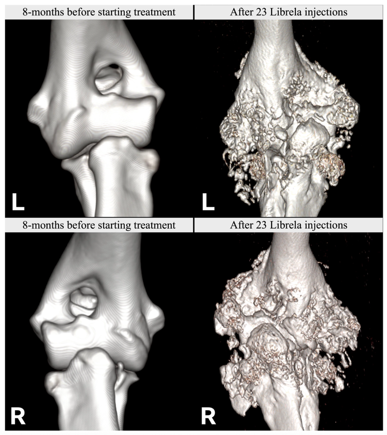
3D CT reconstructions of both elbow joints: 8 months before treatment versus after 23 Librela injections. Catastrophic bilateral remodelling of bony architecture.
Figure 7. 3D CT volume reconstructions of both elbow joints. Top left (L and R): 8 months before starting Librela therapy — visible degenerative changes, but preserved basic joint architecture. Bottom right (L and R): After 23 monthly Librela injections — catastrophic remodelling of the entire bony architecture with extensive periosteal reaction, moth-eaten bone destruction, and complete loss of normal joint contour bilaterally. The appearance is consistent with Rapidly Progressive Osteoarthritis (RPOA) as described in human medicine under anti-NGF therapy. Source: Farrell M et al. Frontiers in Veterinary Science 2025; doi: 10.3389/fvets.2025.1581490

Evidence-based positioning in the OA treatment cascade

Librela should not be first-line or early-line therapy. It should only be considered when all of the following criteria are met:

A. Confirmed end-stage osteoarthritis

  • Clinical OA confirmed by orthopaedic examination
  • Radiographic evidence of advanced degenerative joint disease
  • Significant functional impairment or pain materially affecting quality of life

B. Failure of conservative OA management

Documented inadequate response or intolerance to:

  • NSAIDs
  • Multimodal analgesia (e.g. NSAID ± gabapentin / amantadine / paracetamol / tramadol / others)
  • Intra-articular treatments (corticosteroids, hyaluronate, etc.)
  • Weight loss
  • Environmental modification
  • Exercise modulation
  • Physiotherapy / rehabilitation
  • Nutraceuticals

C. Surgical disease addressed or excluded

  • Surgical salvage performed (arthrodesis / arthroplasty / TPLO etc.), declined by owner, or medically impossible
  • Librela must not replace indicated surgery
  • Librela is not to be used in post-operative pain management

Owners must be explicitly informed that:

  • Severe, rapid, irreversible joint destruction has been reported
  • Pain relief does not equal disease control

Commercial context

The monoclonal antibody franchise for osteoarthritis pain — Librela and Solensia — reached a global value of $568 million in 2025, representing approximately 6% of Zoetis’ total annual revenue. In Q4 2025, the franchise recorded an 11% decline in operating revenue. Zoetis management attributed this primarily to “misinterpretations and influences on social media” regarding Librela’s safety profile.

Despite the revenue decline, Zoetis is doubling down on this technology, having secured approvals for Lenivia and Portela (long-acting versions). The FDA has received adverse event reports on Librela and litigation activity continues, including a shareholder class action alleging Zoetis understated how safety concerns were affecting sales.

Conclusion

Librela is a potent medication that should be reserved as a late-cascade option for dogs with end-stage OA — after failure of conservative multimodal management, and only when surgical solutions are not feasible. If used liberally, there is real risk of significant morbidity. It should be used cautiously.

Please feel free to get in touch with any OA- or Librela-related questions.

Dr. vet. med. Matt Matiasovic MSc Dipl.ECVS

Dr. vet. med. Matt Matiasovic MSc Dipl.ECVS

Small Animal Surgeon · Basel Region