
Arthroscopy in Small Animals: Joint Examination with Minimal Invasiveness
In brief
Arthroscopy in dogs and cats – minimally invasive joint examination with direct visualisation of cartilage, menisci and ligaments. Indications and procedure explained.
Arthroscopy allows direct visual assessment of joint structures with minimal surgical trauma. Compared with conventional open arthrotomy, it offers decisive advantages: less postoperative pain, faster recovery and significantly better visualisation of cartilage surfaces, menisci and ligament structures.
Indications
In dogs and cats, arthroscopic procedures are used primarily for:
- Shoulder joint: Osteochondrosis dissecans (OCD), biceps tendon pathologies
- Elbow joint: Fragmented coronoid process, OCD, elbow dysplasia
- Stifle joint: Meniscal evaluation, cruciate ligament rupture
- Tarsal joint: OCD, cartilage damage
Nanoscopy
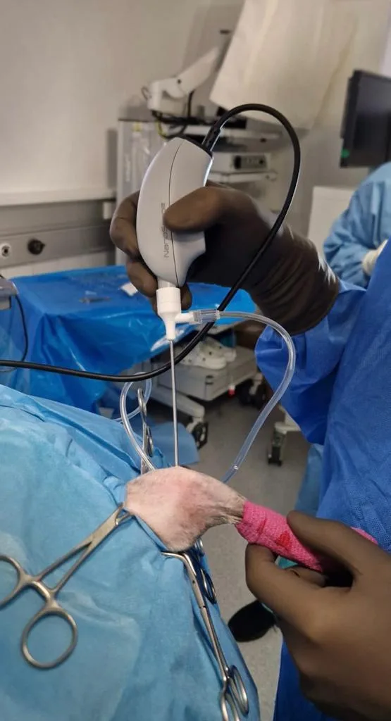
Even less invasive is nano-arthroscopy – here arthroscopes with a diameter of 1.2–1.9 mm are used. These allow arthroscopic examination in locations where conventional arthroscopes would be too large – for example the tarsal joint of cats or small dog breeds.
Clinical Procedure
The procedure is performed under general anaesthesia. After positioning and sterile draping, the arthroscope (camera) is introduced into the joint through a small incision. Visualisation and lavage of the joint, as well as removal of free fragments (for example in elbow dysplasia), can be performed directly arthroscopically.
Recovery time is significantly shorter compared with open surgery – many patients begin bearing weight on the operated limb on the day of surgery.
Summary
Arthroscopy is today an established part of modern small animal orthopaedics. It gives us the ability to perform precise diagnostics and targeted therapy of joint conditions.